
Vol 9 No 1 2024-49
2024..09.01.49
Histopathological effects of Cryptococcus neoformans on liver and kidney in mice

Sara Saad Hussamaldeen Al-Bakir1 and Dalia Abdalkareem Abdalshaheed2*
1Department of Microbiology, College of Veterinary Medicine, University of Baghdad, Baghdad, Iraq; saadsara434@gmail.com.
2Department of Microbiology, College of Veterinary Medicine, University of Baghdad, Baghdad, Iraq; da-lia@covm.uobaghdad.edu.iq.
* Correspondence: dalia@covm.uobaghdad.edu.iq.
Available from. http://dx.doi.org/10.21931/RB/2024.09.01.49
ABSTRACT
This study provides a brief review of approaches for the detection of histopathological effects of Cryptococcus neoformans on the liver and kidney in mice that were injected I/P with 105 yeast cells of C. neoformans suspended in 1 ml phosphate-buffered saline at a single dose. After 14 days, the mice were sacrificed, and histopathological sections from the liver and kidney were prepared and stained with Haematoxylin and Eosin by the P.A.S. method. The results show that the liver was infiltrated with inflammatory cells, primarily mononuclear cells, in the portal. In addition to the activation of Kupffer cells and vacillation of hepatocytes, most blood vessels were congested. The section of the kidney shows sluffing of epithelia lining tubules and destruction of glomeruli, in addition to infiltration of mononuclear cells. These results suggested that the fungus invasiveness of mice has substantial effects on vital organs and may lead to death.
Keywords: Cryptococcus neoformans, Hepatic cryptococcal infection, Cryptococcus.
INTRODUCTION
A fungal illness known as cryptococcosis is brought on by the pathogens Cryptococcus neoformans and C. gattii, which resemble yeast 1. Cryptococcus neoformans serotype A accounts for around 95% of reported cryptococcal infections; the remaining 5% are caused by other serotypes or Cryptococcus gattii 2. It is an illness contracted by inhaling spores or yeast cells that have been dried from environmental sources such as plant matter, soil, and bird excrement 3.
Cryptococcus spp. is widespread and the most significant species concerning medicine 4. When seen under a microscope, the fungus appears as an oval or globular yeast with a diameter of 3mm to 8 mm. It is often encased in a mucopolysaccharidal capsule. The phenoloxidase enzyme causes the capsule to create a significant amount of melanin. The abundance of substrates for phenoloxidase activity in brain tissue may help to partially explain Cryptococcus’s preference for the central nervous system 5. As an encapsulated yeast known as Torulahistolytica or European blastomycosis, Cryptococcus spp. may avoid the immune system’s defense mechanisms and spread primarily from the lungs and central nervous system to the blood, skin, eyes, skeletal system, and urinary tract 7. Cryptococcosis is brought on by inhaling spores or dried yeast cells, and it results in approximately 180,000 fatalities globally each year, including nearly 15% of all AIDS-related deaths 8.
Although a population of C. neoformans may remain dormant for long periods in immunocompetent persons, this often results in an asymptomatic lung infection managed by the host immune response 9. The disease progression results in a highly lethal form of meningoencephalitis 8. Cryptococcosis in animals is a systemic fungal infection of worldwide significance that usually initially infects the nasal cavity, paranasal tissues, or lungs. It can then disseminate to the skin, eyes, or central nervous system. Nasal cryptococcosis is frequently seen in clinical signs, including sneezing, snoring or snorting, dyspnea, nasal deformities, or a mucopurulent, serous, or serosanguineous nasal discharge 10,11. Cryptococcus infections have been reported in many animals, including cats, dogs, horses, birds, and koala bears 12. The most common symptom of hepatic cryptococcal infection is cholestatic jaundice, which can quickly proceed to liver failure and death in cases of widespread illness 13. Cryptococcosis is a relatively uncommon presentation in human patients in urinary tract infections or a diffused illness manifestation (U.T.I.). Patients frequently have an underlying, immune-compromising illness when cryptococcosis is diagnosed in individuals 14. Renal involvement with cryptococcosis in animals is rare, and it has only sporadically been demonstrated in cats with systemic cryptococcosis by detecting fungi during necropsy or urine sediment analysis 15. Therefore, this research aimed to study the histopathological effects of C. neoformanson on the liver and kidney.
MATERIALS AND METHODS
Animals utilized in the experiment
Twenty albino BALB/C female mice weighing an average of 22–25g were used. Animals were housed in cages in pairs and were fed water. A total of 10 mice from the first group (G1) were utilized as the control group and not given any treatment, whereas 10 mice were injected intraperitoneally with a single dose of Cryptococcus neoformans suspension that contained 105 yeast cells into each 1 ml of PBS.
Histopathological sections
Liver and kidney samples were collected after scarification of mice at 14 days of a single administration of C. neoformans and kept in 10% formaldehyde solution for fixation to preserve the figures, size, and tissue for specimens, then processed routinely using the stockinette 16. The specimens were washed with distilled water several times to remove a large proportion of fixative and dehydration by passing the specimens through ascending gradual of ethanol (50,70,80,95 and 100) % in each treatment run with methyl benzoate 24 hrs. Then, they were rehydrated by gradual ethanol (100, 95, 80, 70, and 50) % in each run. The samples were cleared by xylol, embedded in paraffin wax at 70ºC, and sectioned by microtome at 5-6 microns of thickness. The slides were mounted and covered with a coverslip using albumin at 56ºC, stained with Eosin and Hematoxylin, and examined under a light microscope at 400X 17, 18.
RESULTS
Microscopic examination of liver sections for the control group revealed the typical architecture of hepatic lobules and sinusoids lined by thin capillaries and surrounded by a portal area composed of a portal vein, portal artery, and bile ductules in the interstitium (Figure 1a). While the liver of mice infected with C. neoformans showed liver with infiltrated of inflammatory cells, primarily mononuclear cells in the portal area, in addition to activation of Kupffer cells and vacillation of hepatocytes, most blood vessels were congested (Figure 1b & c).

Figure 1: Histopathological section of liver of (a) Control group showing many lobules each lobule contains central vein surrounded by hepatic cord and separated by sinusoid; (b & c) Infected mice with C. neoformans showing infiltration of inflammatory cells, primarily mononuclear cells in the portal area in addition to activation of Kupffer cells and vacillation of hepatocytes, most blood vessels were congested, (H & E stain, 400X).
For the kidney, a microscopic examination of a histological section of control mice showed normal renal tubules. The cortex and normal glomerular tufts were also covered by a thin, dense connective tissue capsule with adipose tissue (Figure 2a). Meanwhile, the kidney section of infected mice with C. neoformansshowsga kidney with sluffing of epithelia lining tubule and complete destruction of glomeruli, in addition to infiltration of mononuclear cells (Figure 2b, c).
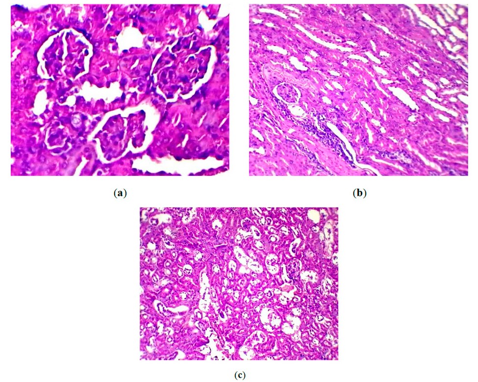
Figure 2: Histopathological section of kidney of (a) Control group showing normal glomeruli and tubules; (b) Infected mice with C. neoformansshowing kidney with sluffing of epithelia lining tubules and destruction of glomeruli with infiltration of mononuclear cells, (H & E stain, 400X).
DISCUSSION
The results showed the hepatic tissue section of mice infected with C. neoformans with vacuolation of hepatocytes, dilation of sinusoids and central veins, as well as portal veins that containing the fibrinous network trapped few P.M.N.s, fibrils of fibrin, precipitated on the endothelial layer of blood vessel cause thickening of the vascular wall and congestion of blood vessels which agreement with Al Kaaby (2009) 19. Additionally, inflammatory cells were infiltrated, especially mononuclear cells, and activation of Kupffer cells in hepatic lobules; fungi can enter the liver or even the whole body through the damaged mucosal membrane, causing aggravated liver damage 20. As observed in figure (2), the renal section of mice treated with C. neoformans revealed destruction of glomerular tuft, sluffing, and convoluted tubules proximal and distal epithelial linings, which are deteriorating with infiltration mononuclear cells. These defects are also seen in another study 21-23. These findings might be due to the dissemination of cryptococcosis. Fungal infection is associated with animals losing weight, their blood cell and leukocyte counts dropping, their plasma glucose levels dropping, and their stomach, liver, and kidneys developing pathological abnormalities 24.
CONCLUSIONS
C. neoformans infection resulted in significant damage to mice’s liver and kidneys. We observed liver damage characterized by vacuolated hepatocytes, dilated blood vessels, and fibrinous network accumulation, suggesting vascular damage, blood congestion, and potential fungal entrapment. Additionally, mononuclear cell infiltration indicated an ongoing immune response. Similarly, kidneys displayed glomerular destruction, epithelial lining disruptions, and mononuclear cell infiltration, suggesting impaired kidney function and potential infection dissemination. These findings align with previous studies and highlight the detrimental effects of C. neoformans on these vital organs. Further research is warranted to explore the specific damage mechanisms and potential treatment options to mitigate organ damage during cryptococcosis infections.
Author Contributions, Conceptualization, S.S.H.A. and D.A.A.; methodology, S.S.H.A.; software, S.S.H.A.; validation, S.S.H.A.; and D.A.A.; formal analysis, S.S.H.A.; investigation, S.S.H.A.; resources, S.S.H.A.; writing-original draft preparation, S.S.H.A.; writing-review and editing, S.S.H.A.; and D.A.A.; visualization, D.A.A.; supervision, D.A.A.; project administration, S.S.H.A.. All authors have read and agreed to the published version of the manuscript.
Funding: This research received no external funding.
Institutional Review Board Statement: The study was conducted according to the guidelines of the Declaration of Helsinki and approved by the Ethics of the Scientific Committee of the Department of Microbiology in the College of Veterinary Medicine, University of Baghdad (Baghdad, Iraq).
Acknowledgments: In this section, you can acknowledge any support given that is not covered by the author’s contribution or funding sections. This may include administrative and technical support or donations in kind (e.g., materials used for experiments).
Conflicts of Interest: The authors declare no conflict of interest.
REFERENCES
1. Kwon-Chung, K.J.; Varma, A. Do major species concepts support one, two or more species within Cryptococcus neoformans? FEMS Yeast Res 2006, 6 (4), 574–587.
2. Maziarz, E.K.; Perfect, J.R. Cryptococcosis. Infect Dis Clin 2016, 30 (1), 179-206.
3. Kwon-Chung, K.J.; Fraser, J.A.; Doering, T.L.; Wang, Z.A.; Janbon, G.; Idnurm, A.; Bahn, Y.S. Cryptococcus neoformansand Cryptococcus gattii, the etiologic agents of cryptococcosis. Cold Spring Harb Perspect Med 2014, 4 (7), 197-216.
4. Zain, H.; Tatar , A.; Alabi, O. M. .; Samiei Zafarghandi, M. . The Effect Of Using Different Vitamin E Levels On The Antioxidants Status Of Broiler Chickens. JLSAR 2023, 4, 37-44.
5. Moretti, M.L.; Resende, M.R.; Lazéra, M.S.; Colombo, A.L.; Shikanai-Yasuda, M.A. Guidelines in cryptococcosis. Rev Soc Bras Med Trop 2008; 41, 524–544.
6. Setianingrum, F.; Rautemaa-Richardson, R.; Denning, D.W. Pulmonary cryptococcosis, a review of pathobiology and clinical aspects. Med Mycol 2019, 57(2),133–150.
7. Kwon-Chung, K.J.; Boekhout, T.; Fell, J.W.; and Diaz, M. Proposal to conserve the name Cryptococcus gattii against C. hondurianus and C. bacillisporus (Basidiomycota, Hymenomycetes, Tremellomycetidae). Taxon 2002, 51(4), 804-806.
8. Rajasingham, R.; Smith, R.M.; Park, B.J.; Jarvis, J.N.; Govender, N.P.; Chiller, T.M.; Denning, D.W.; Loyse, A.; Boulware,D.R. Global burden of disease of HIV-associated cryptococcal meningitis, an updated analysis. Lancet Infect Dis 2017, 17,873–881.
9. A. Al-Badawi, S., T. Al-Wasity, R. AN .Economic Analysis Of The Most Important Variables Affecting Agricultural Employment In Iraq For The Period (1998 – 2019). Anbar Journal Of Agricultural Sciences, 2023; 21(1): 224-249. doi: 10.32649/ajas.2023.179764.
10. Meng, H.C.; Lu, C.H.; Wang, H.C.; Chen, H.L.; Tsai, N.W.; Li, S.H.; Hsu, N.W.; Lin, W.M.; Kung, C.T.; and Lin, W.C. Long-Term Neuropsychological Sequelae in HIVSeronegativeCryptococcal Meningoencephalitis Patients with and without Ventriculoperitoneal Shunts, A Cine M.R.I. Study. Behav Neurol 2015, 1-10.
11. Pimenta, P.; Sofia, A.; João, B.; Maria, J.; Pereira, L.; Paula Maduro, A.; LuísCardoso, C.C. Blepharitis due to Cryptococcus neoformans in a cat from northern Portugal. J Feline Med Surg 2015, 1, 20-25.
12. Malik, R.; Krockenberger, M.B.; Brien, C.R.O.; Carter, D.E.E.A.; Meyer, W.; Canfield, P.J. Veterinary insights into cryptococcosis caused by Cryptococcus neoformans and Cryptococcus gattii. In, Heitman J, Kozel T, Kwon-ChungKJ, Perfect JR, Casadevall A, eds. Cryptococcus, From Human Pathogen to Model Yeast. Washington, DC, A.S.M. Press, 2011, 489–502.
13. A A Al-Azzami , Th T Mohammed . Effect of Adding Dry Leaves of Lemongrass (Cymbopogon Citratus) To the Diet on Some Biochemical Tests of Blood in Broiler (Ross 308). I.O.P. Conf Ser Earth Environ Sci 2023, 1252 (1), 12125. https://doi.org/10.1088/1755-1315/1252/1/012125.
14. Kiertiburanakul, S.; Sungkanuparph, S.; Buabut, B. Cryptococcuria as a manifestation of disseminated cryptococcosis and isolated urinary tract infection. Jpn J Infect Dis 2004, 57, 203-205.
15. Malik, R.; Krockenberger, M.; O’Brien, C. Cryptococcosis. In, Greene CE, ed. Infectious Disease of the Dog and Cat. 3rd ed. Missouri, Saunders, Elsevier 2006,584-598.
16. Luna, L.E. Vegetalismo, shamanism among the mestizo population of the Peruvian Amazon. Vol. 27, Stockholm, Almqvist&Wiksell International, 1986. Pp,429.
17. Ameen M. Shaman , Th. T. Mohammed. Effect Using Feed Additives Instead of Imported Premixes Affects the Physiology of Broiler Chickens. I.O.P. Conf Ser Earth Environ Sci 2023, 1262 (7), 72080. https://doi.org/10.1088/1755-1315/1262/7/072080.
18. Elkhateeb, S. Z.; Ebraheem, M. O.; Abdulateef, S. M.; Ahmed, I. A. Constraints Affecting the Welfare of Domestic Sheep Grazing in the Natural Pasture. I.O.P. Conf Ser Earth Environ Sci 2023, 1252 (1), 12144. https://doi.org/10.1088/1755-1315/1252/1/012144.
19. Al-Shaeli, S.J.; Ethaeb, A.M.; Gharban, H.A. Molecular and histopathological identification of ovine neosporosis (Neospora caninum) in aborted ewes in Iraq. Vet World 2020, 13(3), 597-603.
20. Al Kaaby, H.T. Isolation of Cryptococcus neofrmans from pigeon dropping and study of some pathogenesis aspects. J Kerbala Univer 2009, 7 (2), 316-321
21. A A Al-Azzami , Th T Mohammed . The Effect of Adding Lemongrass Leaf Powder (Cymbopogon Citratus) to the Diet as a Natural Supplement on Some Productive Traits and Oxidation Indicators in Broiler (Ross 308). I.O.P. Conf Ser Earth Environ Sci 2023, 1252 (1), 12123. https://doi.org/10.1088/1755-1315/1252/1/012123.
22. Ramdial, P.K.; Sing Y.; Deonarain, J.; Bhimma, R.; Chotey, N.; Sewram, V. Pediatric renal cryptococcosis, novel manifestations in the acquired immunodeficiency syndrome era. Int J Surg Pathol 2011, 19, 386–392.
23. Pongmekin, P.; Chongtrakool, P.; Santanirand, P.; Kiertiburanakul, S. Clinical characteristics and mortality risk factors of cryptococcal infection among HIV-negative patients. J Med Assoc Thai 2014, 97,36-43.
24. Abbas, O.S.; Abdualshahed, D.A. Effect of Grape Seed Extract on the T-2 Toxicity in mice. Iraqi J Cancer Med Gene 2013, 6(2), 120-125.
Received: October 9th 2023/ Accepted: January 15th 2024 / Published:15 February 2024
Citation: Al-Bakir, S.S.H.; Abdalshaheed, D.A. Histopathological effects of Cryptococcus neoformans on liver and kidney in mice. Revis Bionatura 2024; 9 (1) 49. http://dx.doi.org/10.21931/RB/2024.09.01.49
Additional information Correspondence should be addressed to dalia@covm.uobaghdad.edu.iq.
Peer review information. Bionatura thanks anonymous reviewer(s) for their contribution to the peer review of this work using https://reviewerlocator.webofscience.com/
All articles published by Bionatura Journal are made freely and permanently accessible online immediately upon publication, without subscription charges or registration barriers.
Bionatura ISSN. First 13909355 Ecuador. Scopus coverage years: from 2016 to the present
Publisher’s Note: Bionatura stays neutral concerning jurisdictional claims in published maps and institutional affiliations.
Copyright: © 2023 by the authors. They were submitted for possible open-access publication under the terms and conditions of the Creative Commons Attribution (CC BY) license (https://creativecommons.org/licenses/by/4.0/).
Vol11 No1 2026

INDEXADA EN
INDEXADA EN
