Vol 8 No 4 2023 – 18
Hasanain Emran 1*, Khyreia Habeeb 2, Abeer Majeed 1
1 Departments of Veterinary Surgery and Obstetrics, College of Veterinary Medicine/ University of Baghdad. Baghdad, Iraq; hassanain.j@covm.uobaghdad.edu.iq.
2 Departments of Anatomy and Histology, College of Veterinary Medicine/ University of Baghdad. Baghdad, Iraq; khayria.k@covm.uobaghdad.edu.iq.
1 Departments of Veterinary Surgery and Obstetrics College of Veterinary Medicine/University of Baghdad. Baghdad, Iraq; abeera@covm.uobaghdad.edu.iq.
* Corresponding author: E-mail: hassanain.j@covm.uobaghdad.edu.iq.
Available from. http://dx.doi.org/10.21931/RB/2023.08.04.19
ABSTRACT
This study assessed the histomorphological changes in testicular tissues due to the experimental diabetes mellitus (DM) generated in rabbits with Alloxan. Twenty-four adult male rabbits were used and allocated randomly into two equal groups (n=12 of each). The first group served as a control group and received normal saline only. The second group (diabetic group) was injected with Alloxan at a 100mg/kg IP dose. The serum glucose level was estimated at (0 min), 3 days and then (1,2 and 3 months), by aspiration of a blood sample from the jugular vein mixed with anticoagulant (EDTA) to obtain serum. The testosterone hormone was assayed from the serum obtained by centrifugation of blood samples using a special kit. Testicular biopsies were harvested through surgical castration at (1,2, and 3 months) post-administration of Alloxan. Results displayed a marked increase in glucose levels in the diabetic group starting from day three till the end of the experiment, with high differences between the two groups.
Moreover, testosterone levels significantly decreased in the diabetic group compared to the control group. Our histomorphological examination confirmed that Alloxan leads to testicular damage in rabbits, represented by testicular atrophy, necrosis focusing in spermatogenic cells and separation of germinal epithelium. In conclusion, the injection of Alloxan succeeded in inducing (DM) with clear alteration in studied parameters.
.
Key Words: Biochemical, Testicular Parameters, Alloxan, Rabbits.
INTRODUCTION
Diabetes Mellitus (DM) is a prevalent chronic metabolic disorder. It is considered one of the significant diseases of our time and should not be neglected due to damage and dysfunction in multiple organ systems. It is now the seventh greatest killer of humankind due to its high prevalence and mortality1. DM is broadly classified under two major categories, which include insulin-dependent DM (type I) due to loss or destruction of β-Langerhans islet cells of the pancreas, which leads to an absolute lack of insulin; this type affects children and young adults and non-insulin-dependent (DM) (type 2) in the adult in which β cells retain some functional ability2. DM is characterized by increased blood sugar levels (hyperglycemia), glycosuria, polyuria, polyphagia, polydipsia, and abnormal lipid and protein metabolism3,4. Furthermore, DM is mostly accompanied by severe complications such as macro-vascular diseases, mainly myocardial infarction, congestive cardiac failure, and arteriosclerosis5, and micro-vascular complications include diabetic neuropathy, diabetic nephropathy, and diabetic retinopathy6. Moreover, several studies indicated that DM has a detrimental effect on male sexual and reproductive functions as it lowers gonadotropins and testosterone hormones (responsible for spermatogenesis), resulting in decreased libido, delayed sexual maturation, and infertility with poor semen quality considering the central role of the testis represented by the production of testosterone and sperm which work to retain the characteristics of male and species preservation 7,8,9.
Experimental induction of DM can be achieved surgically through total or partial pancreatectomy10. The pharmacological induction of DM can be achieved by injecting chemical agents such as Alloxan and Streptozotocin, which are most commonly used to cause damage to β cells of the pancreas as their diabetogenic dose is usually 4 to 5 times less than their lethal dose. The dose of these agents varies among the animal species, age, and nutritional status. It can be administered IV, IP, or SC. 11. The present study aimed to assess the histomorphological changes in testicular tissues in diabetic rabbits to identify potential factors that will help design more targeted experiments.
MATERIALS AND METHODS
Experimental animals
The current study was conducted on (24) normoglycemic local strain male rabbits, with an average age ranging from 12-16 months and initial weight ranging from 2000g to 2500g. The rabbits were obtained from the animal house of the faculty of Veterinary Medicine/University of Baghdad.
Management of the animals
The rabbits were kept in individual standard cages under the same managerial and hygienic conditions for three weeks before diabetes induction in the animal’s house at the College of Veterinary Medicine/ University of Baghdad to become familiar with their new home and to monitor any possible abnormalities that may have happened. The cages were floored with a layer of sawdust for rabbits’ urine absorption due to excess urination of rabbits after Alloxan injection. Rabbits were maintained under well-ventilated conditions with 14 hrs. light/ day, controlled temperature (23-25Co), and humidity (40- 45). They were fed standard pellets and greens feed (3 times a day) and were offered to tap water ad libtum throughout the experimental period. During quarantine, rabbits were screened and dewormed with a prophylactic anthelminthic drug (Ivomec1%, 0.2 mg/kg SC; India) against endo and ectoparasites. All works were conducted at the same time in the morning to avoid the effect of daily rhythm on the study results. One day before study initiation, rabbits were weighed using an electronic balance scale (China). The experimental protocols used in the current study were approved by the Institutional Animal Ethics Committee, Faculty of Veterinary Sciences and Animal Husbandry, and conform to the guidelines for the Care and Use of Laboratory Animals for Research. The duration of the experiment was set at three months.
Induction of Diabetes Mellitus
Rabbits (n=24) were used, and they fasted overnight before Alloxan administration; water was not withdrawn before injection of Alloxan. The ventral lateral abdominal area (lateral to the umbilicus) was prepared aseptically before injection under mild sedation (Xylazine hydrochloride 2%, 3 mg/kg BW IM; Spain). 12. A fresh solution of Alloxan monohydrate (Sigma, Germany) was prepared by dissolving it in 0.9% NaCl saline in a concentration of 10% and used immediately (since it is known to be very unstable). The rabbits were randomly and equally divided into two groups. The first group (n=12) was a standard control group, injected with only normal saline (IP) as a vehicle in a 1ml/kg BW dose. The second group (n=12) served as the Alloxaned diabetic group, in which diabetes was induced by injection of 100 mg/kg BW, Alloxan (IP) after dissolved in isotonic solution13, and the research was focused on this group. Before giving Alloxan, the average blood glucose levels of all rabbits were estimated. Immediately after two hours from alloxan administration, rabbits were given 20 ml of 5% glucose solution to counteract the early phase of drug-induced hypoglycemia shock. The blood glucose levels were measured three days post-Alloxan administration using a manual glucometer (Accu-Chech Active, Roche Diagnostics, Germany). Rabbits with blood glucose more than 200 mg/dl were declared diabetic and were selected for the study.
Study parameters
For estimation of glucose levels, three milliliters of blood samples were drawn from each rabbit through jugular vein puncture under aseptic condition at zero time ((base data) and then (3 days, one, two, and three months) post-Alloxan injection for control and diabetic groups. The samples were put into labeled centrifuge tubes containing anticoagulant Ethylene-Diamine-Tetra-Acetic acid (EDTA), then centrifuged at 4000 revolutions per minute for 10 minutes to recover blood serum and stored under refrigerated condition before glucose analysis was carried out. The glucose values were recorded immediately as mg/dl.
The testosterone assay (ng/ml) was performed by collecting venous blood samples from all rabbits in the morning before access to feed and water. Sera were removed by centrifuging blood at 4000 revolutions per minute for 10 minutes and then stored at -20 C° until assayed. The serum testosterone hormone concentration was measured by radioimmunoassay (RIA) using commercial kits obtained from (Diagnostic Systems Laboratories Inc. Texas, USA). This hormone was measured before Alloxan injection (zero time) and then (one, two, and three months) post-Alloxan injection for control and diabetic groups.
Histopathological assessments of testes
All rabbits were subjected to surgical castration (orchidectomy) under the effect of mild general anesthesia represented by Medetomidine (1mg/kg) and Ketamine (100 mg/ml) combination in a dose of 0.25 mg/kg and 30 mg/kg, respectively and administered IM14,15. Testicular tissue biopsies were examined monthly (for three months) (12 bucks/group) and (4 bucks/period). The biopsies were harvested and carefully trimmed to a suitable size (1cm3), fixed in bouin solution for 24 hrs. The samples were labeled and washed in running tap water before processing and then transferred to a graded series of ethanol (70%, 80%, 90%, and 100%), cleared in xylene. The tissues were infiltrated in molten paraffin wax in an oven at 58°C., after which the tissues were embedded in wax and blocked out. The paraffin-embedded biopsies were sectioned at 5μm thickness using a rotatory microtome (Semi-automated Rotary Microtome Wetzlar, Germany). The histological section was stained with Hematoxylin and Eosin (H & E)16 and observed thoroughly under a light microscope equipped with a camera (Olympus, Tokyo, Japan).
Statistical Analysis
All data generated were reported as (Mean ± SEM). In addition, Way Analysis of Variance (ANOVA) was used to test differences between the two groups, followed by Duncan’s Multiple Range test. The significance level was set at P<0.0517.
RESULTS
Clinical signs
Three days post-Alloxan injection, there were clear signs of hyperglycemia as observed in the diabetic rabbit’s group, represented by frequent urination, excessive thirst, decreased physical activity, and a tendency to lie down compared to the control rabbits group, reflecting standard behavioral patterns.
Estimation of serum glucose concentration
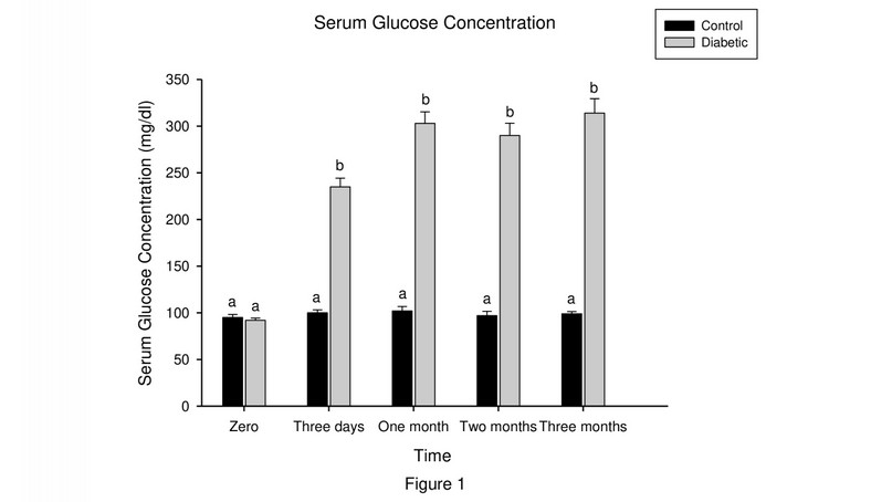
In Alloxan-induced diabetic rabbits, glucose concentration increased steadily (P<0.05) up to three months with the highest value (314±15.33 mg/dl) at this time compared to control group rabbits, which remained relatively stable (standard level) during all periods of observations at about (95 and102 mg/dl). Generally speaking, there were significant differences (P<0.05) between both groups starting from day three and forward periods (Figure 1).

Figure 1. Mean serum glucose values (mg/dl) in two groups at different intervals. No.=12 rabbits/group. Means with different vertical superscripts represent significant differences at (P<0.05) between the control and diabetic groups.
Serum testosterone assay
The testosterone values showed no significant (P> 0.05) difference between the control and diabetic groups at zero time. In contrast, testosterone levels decreased remarkably (p<0.05) in the diabetic rabbits group with the advancement of experimental time. It was recorded (0.09±0.00ng/ml) at the end of the experiment compared to the control group (0.67±0.15ng/ml) (Figure 2).

Figure 2. Mean testosterone values (ng/ml) in two groups at different intervals. No.=12 rabbits/group. Means with different vertical superscripts represent a significant difference (P<0.05) between the control and diabetic groups.
Histopathological findings
Control group
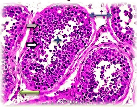
Photomicrographs in control testes depict normal histo-architecture of the testes, which expresses Seminiferous Tubule (ST), which had a definite tubular basement membrane and lined by germ cells epithelium containing different types of germ cells; Spermatogonia lying on the basement membrane beneath it is the supporting Sertoli cells. The interstitial tissues found between ST contain interstitial cells or Leydig cells (Figure 3).

Figure 3. Histology in the testis of the control group shows ST lined by a basement membrane (blue arrow) containing spermatogonia (gray arrow) and Sertoli cells (black arrow), different stages of spermatozoa in the lumen (star) and interstitial tissue between the tubules containing Leydig cells (green arrow) (H&E; X400).
Diabetic group
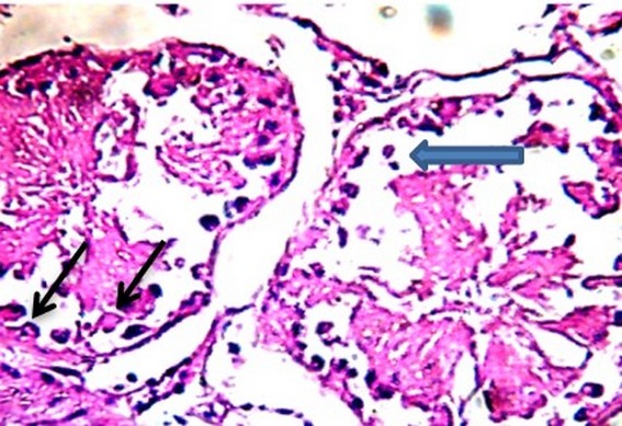
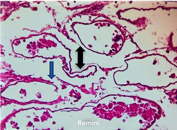
Photomicrograph of the testis, one-month post-injection of Alloxan, showed atrophy of ST, which contains degenerated germ cells, thrombosis of tunica vasculosa with fibrosis of tunica albuginea and intertubular tissue (Figure 4). At two months, the testicular section revealed a decrease in epithelial cells of the ST and vacuolation of Sertoli cells (Figure 5). In another section, there was vacuolar degeneration of spermatogonia with the formation of intratubular multinucleated spermatid giant cells (Figure 6). At the end of the experiment (three months), the testicular section displayed necrosis of all ST with mononuclear cell infiltration around the remnant tubules (Figure 7), in addition to the configuration of ST, which devoided sperms, destruction, and sloughing of the basement membrane (Figure 8).

Figure 4. Photomicrograph in the testis of the diabetic group, one-month post-injection of Alloxan, show atrophy of ST, which contain degenerated germ cells (black arrow), thrombosis of tunica vasculosa (blue arrow) with fibrosis of tunica albuginea (green arrow) and intertubular tissue (red arrow) (H&E; X400).

Figure 5. Photomicrographs in the testis of the diabetic group, two months post-injection of Alloxan, show a decrease in epithelial cells of the ST (blue arrows) and vacuolation of Sertoli cells (black arrows). (H&E; X400).

Figure 6. Photomicrograph in the testis of the diabetic group, two months post-injection of Alloxan, show vacuolar degeneration of spermatogonia (black arrows) with formation of intratubular multinucleated spermatid giant cell (blue arrow). (H&E; X400).

Figure 7. Photomicrographs in the testis of the diabetic group, three months post-injection of Alloxan, show necrosis of all ST (black arrows) with mononuclear cell infiltration around the remnant ST (blue arrow). (H&E; X400).

Figure 8. Photomicrograph in the testis of the diabetic group, three-month post-injection of Alloxan, shows the configuration of ST, which devoided sperms (black arrow), destruction and sloughing of the basement membrane (blue arrow) (H&E; X400).
DISCUSSION
The results of the present study declared that IP injection of a single dose of Alloxan induced a model of DM in rabbits. After three days, the hallmark feature was persistent hyperglycemia, which was monitored throughout the experiment in the diabetic group. These might be attributed to the specific, irreversible cytotoxic effect of Alloxan, which rapidly damages the β cells of the islets of Langerhans in the pancreas and subsequently results in a sharp reduction of insulin secretion from these cells and impairment of glucose uptake by muscle and fat cells, this interpretation was support by18 who reported that apparent increase in glucose level in Alloxan-induced diabetic rabbits was attributed to marked damage to β cells and reducing insulin secretion resulting in hyperglycemia. On the other hand, previous study 19 suggested that Alloxan and its metabolites tend to concentrate in pancreatic islet tissue relative to some other tissue. The harmful effects of Alloxan, causing hyperglycemia, might be due to rapid inhibition of the insulin secretory mechanism.
Furthermore, 20 observed that glucose formation is promoted in insulin deficiency, partly due to increased conversion of glycogen to glucose but mainly by gluconeogenesis. Similarly, in an experimental trial in mice, Syiem reported that Alloxan is the most prominent diabetogenic chemical used in diabetes research21. It is a cytotoxic glucose analogue causing selective destruction of β-cells in the pancreas through the generation of reactive oxygen system (ROS), which causes severe anemia responsible for hyperglycemia and many other complications.
The clinical signs of hyperglycemia, as observed in studied rabbits, were constant with that noticed by 22when they induced diabetes in the dog at the same dose of Alloxan used in our study, which included polyuria, polyphagia, glycosuria, in-appetence, general weakness, depression and recumbency. Likewise, 23 recorded polydipsia, polyuria, and polyphagia in diabetic rats.
In the present work, it is noteworthy herein to mention that serum testosterone level was remarkably decreased in the diabetic group, which is one of the indicators of dysfunction in testicular androgen metabolism; this may be due to the diminished number of Leydig cells, which are responsible for the secretion of this hormone, evoked from the harmful effect of DM. This interpretation is fundamentally consistent with a previous study by 24 who noticed a decrease in testicular testosterone, which is associated with histological alterations in androgen target cells, i.e., Leydig cells. In an experimental trial, 25 have reported that hyperglycemia causes dysfunction of Leydig cells. Thus, these cells cannot secrete testosterone, which can cause a reduction of serum levels of this main androgenic hormone level in streptozotocin-induced diabetes in male rats.
The testis is a vital organ in the male reproductive system and is well vascularized; it is located in an «out-pocketing» of the abdominal cavity called the scrotum. Testes have dual functions, i.e., exocrine (spermatogenesis) and endocrine (synthesis of male hormones, mainly testosterone). Histologically, the testes are enclosed in a testicular tissue capsule through which blood vessels and nerves arrive and depart from the organ. The testes comprise three layers: tunica albuginea, tunica vaginalis, and tunica vasculosa26.
The histological section of the normal testis in the present investigation showed cells, including spermatogonia and Sertoli cells, which were seen laying on the basement membrane of ST in addition to different stages of spermatozoa. The Leydig cells were present in between the tubules.
In our study, the diabetic group reflected atrophy of ST with aggregations of tissue debris representing dead germ cells in their lumen. Furthermore, vacuolation of Sertoli cells is a common feature of morphological injury of Sertoli cells and often accompanies generalized germinal cell degeneration/atrophy. This was indicative of the harmful effect of Alloxan. The present findings were in agreement with a study in rat27.
Moreover, we noticed multi-core giant cells in the ST. Similarly, Cernochova and Kamarad observed multinucleated giant cells in some ST. At the same time, these cells were found in systemic, toxic, infectious, ischemic pathologies and in cryptorchidism, causing tubular atrophy28.
Other histopathological sections in the current study in the diabetic group revealed an irregular shape of ST, which was devoid of germ cells, absence of Leydig cells, which was replaced by fibrous connective tissue containing mononuclear cells infiltration in addition to increased sloughing of germinal epithelium. This may be due to a lack of testosterone, which responds to the spermatogenesis process and finally causes histopathological damage in the testis. Monsees suggested that the decreased testosterone reflected on Sertoli cells function leads to loss of germ cells, loss of contact between them, reduction of the thickness of the seminiferous epithelium, and finally, destruction of testicular tissue29. Moreover, it was speculated that the pathological changes of seminiferous epithelium may disrupt the Sertoli and germ cells and, therefore, spermatogenesis and may also lead to germ cell loss30. Elshennawy and Abo Elwafa 31reported that spermatogonia are highly sensitive to toxicants because of their mitotic activity.
Moreover, the defects of spermatogonia will affect the development of the stages of spermatogenesis. Also, the present results declared that ST has various degrees of degeneration of spermatogonia with numerous apoptotic cells. These findings are in agreement with that were reported by Al‑Damegh32.
CONCLUSION
In conclusion, the results of the current study reveal that DM in rabbits was successfully induced with a single dose of Alloxan via the IP route. It caused a marked elevation in serum glucose levels and a severely diminution of testosterone values. Furthermore, DM alters histomorphological parameters of testicles like testicular atrophy and degenerative changes in the testicles, which were prominent especially at the end of the experiment in the diabetic group compared to the control group and reduces sperm production in an animal model.
REFERENCES
1. Jerrold, M.O. Prospects for research in DM. J. Am. Med. Assoc., 2001; 285:628-632.
2. Burski, K., Ueland, T., Maciejewski, R. Serum amylase activity disorders in the course of experimental diabetes in rabbits. Vet. Med. Czech., 2004; 49:197-200.
3. Wong, J., Molyneaux, L., Constantio, M.I., Twing, S.M., Yue, DK. Metabolic syndrome in type 2 diabetes: Diabet. Obes. Metabol., 2006; 8:690-697.
4. Bakýrel, T., Bakýrel, U., Yardibi, H. In vivo assessment of anti-diabetic and antioxidant activities in Alloxan-diabetic rabbits. J Ethnopharmaco., 2008; 116: 64-73.
5. Hyvorinen, M., Qiao, Q., Tuomilehto, J., Laatikainen, T., Heine, R., Stegmayr, B. Hyperglycemia and stroke mortality: comparison between fasting and 2-hrs glucose criteria. Diabetes Care, 2009; 32:348-354.
6. Basit, A., Hydrie, M. Z., Hakeem, R., Masood, Q. Frequency of chronic complications of type II diabetes. J. Coll. Physicians Surg. Pak., 2004; 14:79-83.
7. Sofikitis, N., Giotitsas, N., Tsounapi, P. Hormonal regulation of spermatogenesis J. Steroid Biochem. Mol. Biol., 2008; 109:323-330.
8. Mohasseb, M., Ebied, S., Yehia, M.A., Hussein, N. Testicular oxidative damage and role of combined antioxidant supplementation in experimental diabetic rats. J. Physiol. biochem., 2011; 67(2): 185-194.
9. Chikaodiri, H. O. Reproductive Physiology of Male Rabbits: A Key Factor in Buck Selection for Breeding. Advances in Reprod. Sci., 2020; 8 (2): 37-42.
10. Misra, M., Aiman, U. Alloxan: An unpredictable drug for diabetes induction? Indian J. Pharmacol., 2012; 44(4): 538-539.
11. Tripathi, V., Verma, J. Different models used to induce diabetes: a comprehensive review. Int. J. Pharm. Sci., 2014; 6 (6):29-32.
12. Camkerten, I., Sindak, N., Ozkurt, G., Ipek, H., Biricik, H. S., Sahin, T. Effect of ketamine-xylazine anesthesia on some hematological and serum biochemical values of Bozova Greyhounds. Veterinary faculty Dergisi Harran University, 2013; 2(1): 27-31.
13. Saadla, S. A., Abdul-Hameed, K., Ghazanfar, A., Saadatull, A. K. Alloxan induced diabetes in rabbits. Pak. J. of Pharmaco., 2005; 22 (2):41-45.
14. Orr, H. E., Roughan, J. V., Flecknell, P. A. Assessment of ketamine and medetomidine anesthesia in rabbits. Vet. Anesth. Analg., 2005; 32: 271-275.
15. Khaleefa, M. A., Emran, H. A. Histopathological study of the effect of using repeated doses of platelet-rich plasma on articular cartilage repair in Rabbits. Iraqi Journal of Veterinary Sciences, 2022; 36(Supplement I): 203-210. doi: 10.33899/ijvs.2022.135928.2543
16. Chan JK. «The wonderful colors of the hematoxylin-eosin stain in diagnostic surgical pathology». Int J Surg Pathol. 2014; 22 (1): 12–32. doi:10.1177/1066896913517939. PMID 24406626.
17. Snedecor, G. W., Cochran, W. G. Statistical Methods 7th Ed. The Stae University Press American, Iowa 1982.
18. Arai, T., Nakamura, M., Magori, E., Fukuda, H., Sako, T. Changes in activities of enzymes related to energy metabolism in peripheral leukocytes of diabetic dogs with glycemic control by intensive insulin treatment. Res. Vet. Sci., 2002; 73: 183-186.
19. Baynes, J.W. Role of oxidative stress in development of complications in diabetes. Diabetes 1991; 40:405-412.
20. Kimmel, S.E., Ward, C.R., Hess, R.S. Familial insulin-dependent diabetes mellitus in dogs. J. Am. Anim. Hospital Assoc., 2002; 38, 235-238.
21. Syiem, D., Syngai, G., Khup, P.Z., Khongwir, B.S., Kharbul, I. B., Kayang, H. Hypoglycemic effects of Potentilla fulgens L in normal and Alloxan- induced diabetic mice. J. Ethnopharmacol., 2002; 83(1-2): 55-61.
22. Kim, J.M., Chung, J.Y., Lee, S.Y., Young, H.Y. Hypoglycemic effects of vanadium on Alloxan induced diabetic dogs. J. Vet. Sci., 2006; 7:391-395.
23. Jelodar, G., Khaksar, Z., Pourahmadi, M. Endocrine profile and testicular histomorphometry at puberty in rat offspring from diabetic mothers. Comparative Clinical Pathology, 2010; 19: 135-139.
24. Yang, Z. W., Kong, L. S., Guo, Y. Histological changes of the testis and epididymis in adult rats as a result of Leydig cells destruction after ethane dimethane sulfonate treatment: a morphometric study. Asian J. of Andrology, 2006; 8: 289-299.
25. Ogunyinka, B. I., Oyinloye, B. E., Kappo, A.P. Modulatory influence of Parkia biglobosa protein isolate on testosterone and biomarkers of oxidative stress in brain and testes of streptozotocin-induced diabetic in male rats. Int. J. Physiol. Pathophysiol. Pharmacol., 2016; 8(3): 78-86.
26. Smith, L.B., Walker, W.H. The regulation of spermatogenesis by androgens. Semin Cell Dev. Biol., 2014; 30:2-13.
27. Anniballo, R., Ubaldi, F., Cobellis, L., Rienzi, L., Greco, E., Tesarik J. Criteria predicting the absence of spermatozoa in the Sertoli cell-only syndrome can be used to improve success rates of sperm retrieval. Hum. Reprod., 2000; 15(11):2269-2277.
28. Cernochova, D., Kamarad, V. Toxic effect of lead of mice testicles after its administration with drinking water, Acta Universitatis Palackianae Olomucensis Facultatis Medicae, 1992; 133: 9-13.
29. Monsees, T. K., Franz, M., Gebhardt, S., Schill, W. B., Hayatpour, J. Sertoli cells as a target for reproductive hazards. Andrologia, 2002; 32:239-46.
30. Xie, B., Li, J., Zhu, W. Pathological changes of testicular tissue in normal adult mice: A retrospective analysis. Exp. Ther. Med., 2014; 7(3): 654-656.
31. Elshennawy, W., Abo El-Wafa, H. Histological changes in mammalian testis under the effect of Hydrocortisone. J. Am. Sci., 2011; 7(9): 38-48.
32. Al‑Damegh, M.A. Rat testicular impairment induced by electromagnetic radiation and the protective effects of the antioxidants vitamins C and E. Clinics (Sao Paulo). 2012; 67:785‑792.
Received: 28 September 2023/ Accepted: 15 November 2023 / Published:15 December 2023
Citation: Emran H, Habeeb K, Majeed A.The histomorphological appearance of the testes in Alloxan-induced diabetic rabbits. Revis Bionatura 2023;8 (4) 19. http://dx.doi.org/10.21931/RB/2023.08.03.19


